


 TR
TR DE
DE
Salivary Gland Stone
Today, we perform the treatment of salivary gland stones, in other words sialolithiasis, by working with very thin endoscopes through the salivary gland channels. The sialendoscopy method, which we have been using and pioneering since 1990 in the world and since 2004 in our country, together with the advancing medical and optical technology, has enabled us to remove the stones of the patients who are thought to be impossible to be removed, and whose glands are generally recommended to be removed by surgery. Our success in removing salivary gland stones with sialendoscopy and assistive techniques is around 85%.
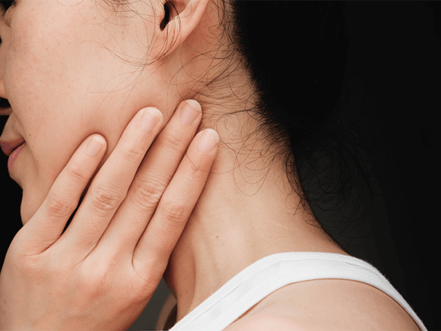
What is Salivary Gland Stone?
The diameter of the salivary ducts is 2-3 mm. The points where they open into the mouth are as large as the needle tip (0.1 mm). Therefore, even very small stones can block the salivary duct; may cause swelling under the cheek or chin. In other words, stones smaller than 2 millimeters or a large stone with a diameter of 3-4 centimeters can cause the patient's salivary gland to swell. In patients whose salivary glands are repeatedly swollen under the chin or cheek, we usually think that there is a stone blocking the salivary duct at first. About 40% of the stones cannot be seen with the films. Today, the most accurate diagnostic method for such conditions is sialendoscopy. No other examination can take the place of sialendoscopy, which allows us to see the salivary gland stone directly.
With diagnostic salivary gland endoscopy, we see what the disease is that obstructs the salivary duct. If this is a stone case, we determine the position of the stone, its dimensions, number and whether it can be removed. In other words, whether any salivary gland stone can be removed can only be understood by seeing it during sialendoscopy. The fact that the stones are large does not mean that they cannot be removed. Because if the stone can be reached completely through the salivary gland duct, we can break the stone and remove it in pieces. In this way, we can remove even very large stones with a diameter of 2.5-3 centimeters. The stone breaking method is required in approximately 80% of cases. Without stone breaking, only stones as small as 3-4 millimeters can be removed by holding them. However, sometimes even small salivary gland stones with a diameter of 2.5-3 millimeters may not be removed without crushing if the stone is stuck in a narrow channel. Small stones can be removed in 5-10 minutes, while large stones may require 4-5 hours of work. Salivary gland endoscopy (sialendoscopy) is the most advanced way of diagnosis and treatment of stenosis in the salivary ducts and other duct diseases, especially sialolithiasis (salivary gland stone).
Salivary Gland Stone Treatment
With interventional sialendoscopy, we can grab and remove stones that are suitable in size and freely circulating in the canal with various tools called forceps and baskets. With these tools, we can remove stones that cannot be held in pieces after breaking them in the canal. The only condition of these methods is to reach the stone through the canal. The success rate in breaking the salivary gland stone is around 80%. The 20% probability of failure depends on some unbreakable stones, salivary gland stones embedded in the duct, or stones in the end branches of the salivary gland (ie inaccessible stones in the gland). However, even in such cases, we can increase the possibility of spontaneous removal of these types of stones over time, as we can perform applications such as widening and shortening the salivary duct during the sialendoscopic approach.
Sialendoscopy is an advanced technological diagnostic method that can be performed by specialists with advanced endoscopy skills and intensive training on sialendoscopy. In order to be able to intervene during sialendoscopy, for example, to perform a treatment such as breaking the salivary gland stone, the physician must be able to use laser or air stone breaking (pneumatic lithotripsy) methods, these devices must be among their equipment and adapted for sialendoscopic use. One of the pioneers of the sialendoscopy method in our country, Dr. You can watch Atilla Şengör's video on the salivary gland stone breaking method from the link below.
Our Clinical Information
He was born in 1981 in Zile district of Tokat. Starting primary school at Rize Atatürk Primary School, Dr. Deniz Yazıcı continued his education life in the Ergani district of Diyarbakır, since his father's place of duty changed since the 4th grade of primary school.